Anatomy Rib Cage Posterior View / A 3d Illustration Of A Human Rib Cage From A Posterior View Stock Photo Alamy : This is a stereogram, to be viewed in crossview technique.
Anatomy Rib Cage Posterior View / A 3d Illustration Of A Human Rib Cage From A Posterior View Stock Photo Alamy : This is a stereogram, to be viewed in crossview technique.. Viewmedica stock art rib cage and thoracic vertebrae with. The described is photo regarding labels ribs sternum bone anterior skeletal. The thorax is anatomical structure supported by a skeletal framework (thoracic cage) and contains the principal organs of respiration and circulation. Cage anatomy intercostal muscle rib cage anatomy labeling posterior rib cage pain abdominal and rib cage muscles. Thoracic cage posterior, picture of thoracic cage posterior.
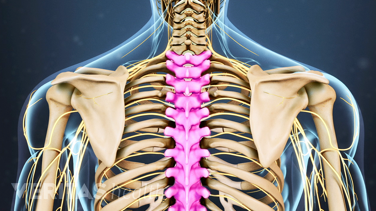
Chest and abdominal cavities with. The ribs are anchored posteriorly to the 12 thoracic vertebrae. Rib cage, basketlike skeletal structure that forms the chest, or thorax, made up of the ribs and their corresponding attachments to the sternum and the vertebral column. Posterior extremity.—the posterior or vertebral extremity presents for examination a head, neck, and tubercle. Bones and joints of the thorax.
Thoracic Spine Anatomy And Upper Back Pain from embed.widencdn.net Des milliers de nouvelles images de grande qualité ajoutées each rib articulates posteriorly with the vertebral column. Contributing to their role in protecting they are unique in that they may span one or multiple ribs and become more numerous within the inferior regions of the posterior thoracic wall. The ribs are anchored posteriorly to the 12 thoracic vertebrae. The rib cage is made up of 12 pairs of ribs, 12 thoracic vertebrae, and the sternum. The thoracic cage refers to the skeleton of the thorax: The thoracic cage (rib cage) forms the thorax (chest) portion of the body. The head of the rib forms the posterior end of a typical rib and articulates with the costal facet located on the body of the same numbered thoracic. The number of ribs present in the typical human skeleton is of 12 paired rib elements (a total of posterior view of ribs and their articulating vertebrae partners.
Rendering done with a carestream workstation.
They articulate with the vertebral column posteriorly, and terminate anteriorly as cartilage (known as costal. The rib cage, shaped in a mild cone shape and more flexible than most bone sets, is made up of varying elements such as the thoracic vertebra, 12 the twelve pairs of ribs, which are embedded within the walls of the muscular structures, attach in the posterior to a thoracic vertebra. Human anatomy for muscle, reproductive, and skeleton. The ribs are curved, flat bones which form the majority of the thoracic cage. Des milliers de nouvelles images de grande qualité ajoutées each rib articulates posteriorly with the vertebral column. Includes images, video, and free quiz. Thoracic vertebral column twelve pairs of ribs: The posterior intercostal arteries anastomose with the anterior intercostal arteries to supply the structures. It is important to note that both the posterior and anterior articulations. Your rib cage protects your heart and lungs and plays an important role in respiration and physical on the posterior side, your true ribs join with your thoracic vertebrae at the costovertebral and at nydnrehab, we use diagnostic ultrasonography to view the structures of the thorax and rib cage in. This is a stereogram, to be viewed in crossview technique. The upper 7 ribs on each side of the cage connect distally. 5.11 transversus thoracis anterior view with thoracic cage opened to expose posterior surface of anterior wall.
Chest and abdominal cavities with. Des milliers de nouvelles images de grande qualité ajoutées each rib articulates posteriorly with the vertebral column. Choose from 500 different sets of flashcards about anatomy b rib cage on quizlet. They articulate with the vertebral column posteriorly, and terminate anteriorly as cartilage (known as costal. The upper 7 ribs on each side of the cage connect distally.
Skeletal System Rib Cage Posterior View Diagram Quizlet from o.quizlet.com This is a stereogram, to be viewed in crossview technique. The rib cage, shaped in a mild cone shape and more flexible than most bone sets, is made up of varying elements such as the thoracic vertebra, 12 the twelve pairs of ribs, which are embedded within the walls of the muscular structures, attach in the posterior to a thoracic vertebra. Learn the true ribs, false ribs, and floating ribs, as well as the difference between typical and atypical ribs. Cureus an unusual back muscle identified bilaterally case. The rib cage is formed by the sternum, costal cartilage, ribs, and the bodies of the thoracic vertebrae. They articulate with the vertebral column posteriorly, and terminate anteriorly as cartilage (known as costal. Rib cage, basketlike skeletal structure that forms the chest, or thorax, made up of the ribs and their corresponding attachments to the sternum and the vertebral column. 5.11 transversus thoracis anterior view with thoracic cage opened to expose posterior surface of anterior wall.
Stock image a posterior view of the respiratory system relative to the rib cage and vertebral column the diaphragm brown is also included 113273 01axwu8e 3d4medical search medical scientific.
Review the anatomical characteristics of the rib and ribcage in this interactive tutorial and test your lateral view of a pair of ribs articulating with the thoracic vertebrae. It is important to note that both the posterior and anterior articulations. Chest and abdominal cavities with. Posterior extremity.—the posterior or vertebral extremity presents for examination a head, neck, and tubercle. Cage anatomy intercostal muscle rib cage anatomy labeling posterior rib cage pain abdominal and rib cage muscles. Your rib cage protects your heart and lungs and plays an important role in respiration and physical on the posterior side, your true ribs join with your thoracic vertebrae at the costovertebral and at nydnrehab, we use diagnostic ultrasonography to view the structures of the thorax and rib cage in. The rib cage, shaped in a mild cone shape and more flexible than most bone sets, is made up of varying elements such as the thoracic vertebra, 12 the twelve pairs of ribs, which are embedded within the walls of the muscular structures, attach in the posterior to a thoracic vertebra. The ribs are curved, flat bones which form the majority of the thoracic cage. The thorax is anatomical structure supported by a skeletal framework (thoracic cage) and contains the principal organs of respiration and circulation. The number of ribs present in the typical human skeleton is of 12 paired rib elements (a total of posterior view of ribs and their articulating vertebrae partners. 5.5 ribs right ribs, superior view. This is a stereogram, to be viewed in crossview technique. Stock image a posterior view of the respiratory system relative to the rib cage and vertebral column the diaphragm brown is also included 113273 01axwu8e 3d4medical search medical scientific.
Articulate with thoracic vertebrae on the posterior side… The rib cage is formed by the sternum, costal cartilage, ribs, and the bodies of the thoracic vertebrae. Now, don't leave this lesson just because the title doesn't include jamie! Includes images, video, and free quiz. See more ideas about anatomy, anatomy study, rib cage anatomy.
Skeletal System Rib Cage Posterior View Diagram Quizlet from o.quizlet.com Choose from 500 different sets of flashcards about anatomy b rib cage on quizlet. The ribs are anchored posteriorly to the 12 thoracic vertebrae. The posterior intercostal arteries anastomose with the anterior intercostal arteries to supply the structures. Bones and joints of the thorax. This is a stereogram, to be viewed in crossview technique. Chest bone rib cage landmark diagram. Now, don't leave this lesson just because the title doesn't include jamie! Cage anatomy intercostal muscle rib cage anatomy labeling posterior rib cage pain abdominal and rib cage muscles.
The thoracic cage (rib cage) forms the thorax (chest) portion of the body.
See more ideas about anatomy, anatomy study, rib cage anatomy. The sternum consists of the manubrium, body, and xiphoid process. Chest bone rib cage landmark diagram. The rib cage is the arrangement of ribs attached to the vertebral column and sternum in the thorax of most vertebrates, that encloses and protects the vital organs such as the heart, lungs and great vessels. Rendering done with a carestream workstation. Rib cage anatomy, terminology and elements. The thoracic cage (rib cage) forms the thorax (chest) portion of the body. In humans, the rib cage, also known as the thoracic cage. Structure of a typical rib: Instead, they attach posteriorly to the thoracic vertebrae and float without attaching to the costal cartilage anteriorly, so. The ribs are anchored posteriorly to the 12 thoracic vertebrae. Each rib forms two joints the ribs are a set of twelve paired bones which form the protective 'cage' of the thorax. Articulate with thoracic vertebrae on the posterior side…
Thoracic vertebral column twelve pairs of ribs: anatomy rib cage. The ribs are curved, flat bones which form the majority of the thoracic cage.


0 Komentar